

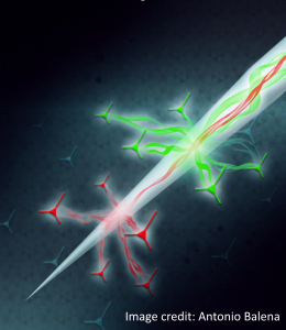
A primary goal of experimental neuroscience is to dissect the neural microcircuitry underlying brain function, ultimately to link specific neural circuits to behavior. There is widespread agreement that innovative new research tools are required to better understand the incredible structural and functional complexity of the brain. To this aim, optical techniques based on genetically encoded neural activity indicators and actuators have represented a revolution for experimental neuroscience, allowing genetic targeting of specific classes of neurons and brain circuits. However, for optical approaches to reach their full potential, we need new generations of devices better able to interface with the extreme complexity and diversity of brain topology and connectivity.
This project aspires to develop innovative technologies for multipoint optical neural interfacing with the mammalian brain in vivo. The limitations of the current state-of-the-art will be surmounted by developing a radically new approach for modal multiplexing and de-multiplexing of light into a single, thin, minimally invasive tapered optical fiber serving as a carrier for multipoint signals to and from the brain. This will be achieved through nano- and micro-structuring of the taper edge, capitalizing on the photonic properties of the tapered waveguide to precisely control light delivery and collection in vivo. This general approach will propel the development of innovative new nano- and micro-photonic devices for studying the living brain.
The main objectives of the proposals are: 1) Development of minimally invasive technologies for versatile, user-defined optogenetic control over deep brain regions; 2) Development of fully integrated high signal-to- noise-ratio optrodes; 3) Development of minimally invasive technologies for multi-point in vivo all-optical “electrophysiology” through a single waveguide; 4) Development of new optical methodologies for dissecting brain circuitry at small and large scale

Plasmonics on a Neural Implant: Engineering Light–Matter Interactions on the Nonplanar Surface of Tapered Optical Fibers
Advanced Optical Materials 10, 2101649 (2022)
Abstract - Optical methods are driving a revolution in neuroscience. Ignited by optogenetic techniques, a set of strategies has emerged to control and monitor neural activity in deep brain regions using implantable photonic probes. A yet unexplored technological leap is exploiting nanoscale light-matter interactions for enhanced bio-sensing, beam-manipulation and opto-thermal heat delivery in the brain. To bridge this gap, we got inspired by the brain cells’ scale to propose a nano-patterned tapered-fiber neural implant featuring highly-curved plasmonic structures (30 μm radius of curvature, sub-50 nm gaps). We describe the nanofabrication process of the probes and characterize their optical properties. We suggest a theoretical framework using the interaction between the guided modes and plasmonic structures to engineer the electric field enhancement at arbitrary depths along the implant, in the visible/near-infrared range. We show that our probes can control the spectral and angular patterns of optical transmission, enhancing the angular emission and collection range beyond the reach of existing optical neural interfaces. Finally, we evaluate the application as fluorescence and Raman probes, with wave-vector selectivity, for multimodal neural applications. We believe our work represents a first step towards a new class of versatile nano-optical neural implants for brain research in health and disease.
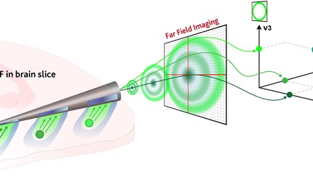
Orthogonalization of far-field detection in tapered optical fibers for depth-selective fiber photometry in brain tissue
APL Photonics 7, 026106 (2022)
Abstract - The field of implantable optical neural interfaces has recently enabled the interrogation of neural circuitry with both cell-type specificity and spatial resolution in sub-cortical structures of the mouse brain. This generated the need to integrate multiple optical channels within the same implantable device, motivating the requirement of multiplexing and demultiplexing techniques. In this article, we present an orthogonalization method of the far-field space to introduce mode-division demultiplexing for collecting fluorescence from the implantable tapered optical fibers. This is achieved by exploiting the correlation between the transversal wavevector kt of the guided light and the position of the fluorescent sources along the implant, an intrinsic property of the taper waveguide. On these bases, we define a basis of orthogonal vectors in the Fourier space, each of which is associated with a depth along the taper, to simultaneously detect and demultiplex the collected signal when the probe is implanted in fixed mouse brain tissue. Our approach complements the existing multiplexing techniques used in silicon-based photonics probes with the advantage of a significant simplification of the probe itself.
Neurophotonic tools for microscopic measurements and manipulation: status report
Neurophotonics 9(S1) 013001 (2022)
Extracted from the text - “We review the current state-of-the-art tools that are, in general, applicable to animal models and usually considered under the umbrella of microscopy. We start with an overview of high-resolution structural imaging tools that have emerged and/or matured within the last decade. Then, we highlight several newly developed optical sensors and modulators of brain activity followed by discussion of a suite of one-photon (1P) and multiphoton fluorescence microscopy methods aimed at large-scale imaging of brain activity with high temporal resolution. We include a number of technologies for imaging cerebral blood flow and metabolism and feature several imaging approaches that integrate optical tools with other measurement modalities providing complementary information and/or helping to overcome fundamental limitations of purely optical methods. Finally, we highlight the importance of computational tools that complement optical technologies extending their capabilities.”
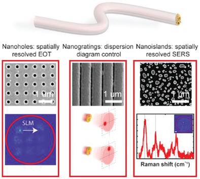
Holographic Manipulation of Nanostructured Fiber Optics Enables Spatially-Resolved, Reconfigurable Optical Control of Plasmonic Local Field Enhancement and SERS
Small
Abstract - Integration of plasmonic structures on step-index optical fibers is attracting interest for both applications and fundamental studies. However, the possibility to dynamically control the coupling between the guided light fields and the plasmonic resonances is hindered by the turbidity of light propagation in multimode fibers (MMFs). This pivotal point strongly limits the range of studies that can benefit from nanostructured fiber optics. Fortunately, harnessing the interaction between plasmonic modes on the fiber tip and the full set of guided modes can bring this technology to a next generation progress. Here, the intrinsic wealth of information of guided modes is exploited to spatiotemporally control the plasmonic resonances of the coupled system. This concept is shown by employing dynamic phase modulation to structure both the response of plasmonic MMFs on the plasmonic facet and their response in the corresponding Fourier plane, achieving spatial selective field enhancement and direct control of the probe's work point in the dispersion diagram. Such a conceptual leap would transform the biomedical applications of holographic endoscopic imaging by integrating new sensing and manipulation capabilities.
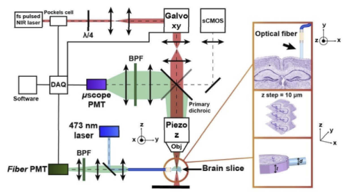
Influence of the anatomical features of different brain regions on the spatial localization of fiber photometry signals
Biomedical Optics Express 12 6081 (2021)
Abstract - Fiber photometry is widely used in neuroscience labs for in vivo detection of functional fluorescence from optical indicators of neuronal activity with a simple optical fiber. The fiber is commonly placed next to the region of interest to both excite and collect the fluorescence signal. However, the path of both excitation and fluorescence photons is altered by the uneven optical properties of the brain, due to local variation of the refractive index, different cellular types, densities and shapes. Nonetheless, the effect of the local anatomy on the actual shape and extent of the volume of tissue that interfaces with the fiber has received little attention so far. To fill this gap, we measured the size and shape of fiber photometry efficiency field in the primary motor and somatosensory cortex, in the hippocampus and in the striatum of the mouse brain, highlighting how their substructures determine the detected signal and the depth at which photons can be mined. Importantly, we show that the information on the spatial expression of the fluorescent probes alone is not sufficient to account for the contribution of local subregions to the overall collected signal, and it must be combined with the optical properties of the tissue adjacent to the fiber tip.